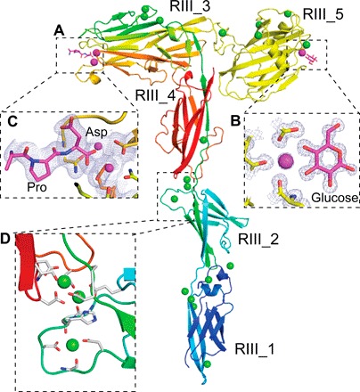
Fig. 4. Detailed structural features of the MpIBP_RIII ligand-binding domains.

(A) RIII_1–4 is colored in rainbow representation, whereas the RIII_5 construct is colored yellow. Calcium ions in the ligand-binding sites are shown as magenta spheres, whereas the other Ca2+ are shown as green spheres. (B) Enlarged view of the sugar-binding site of the RIII_5 structure, showing the 1 Å 2Fo − Fc map and the carbon atoms of the glucose molecule colored magenta. Oxygen atoms are red, and nitrogen atoms are blue. (C) Enlarged view of the ligand-binding cavity of RIII_3 is shown with the 2.1 Å resolution 2Fo − Fc map contoured at 1 σ [as in (B)]. Ca2+ coordination by the C-terminal Pro and Asp residues from a symmetry-related molecule are shown in stick representations. (D) Enlarged view of the Ca2+-stiffened linker region between RIII_2 and RIII_4. Ca2+ coordinating residues are shown in stick representation.
