. 2017 Aug 9;6:e26640. doi: 10.7554/eLife.26640

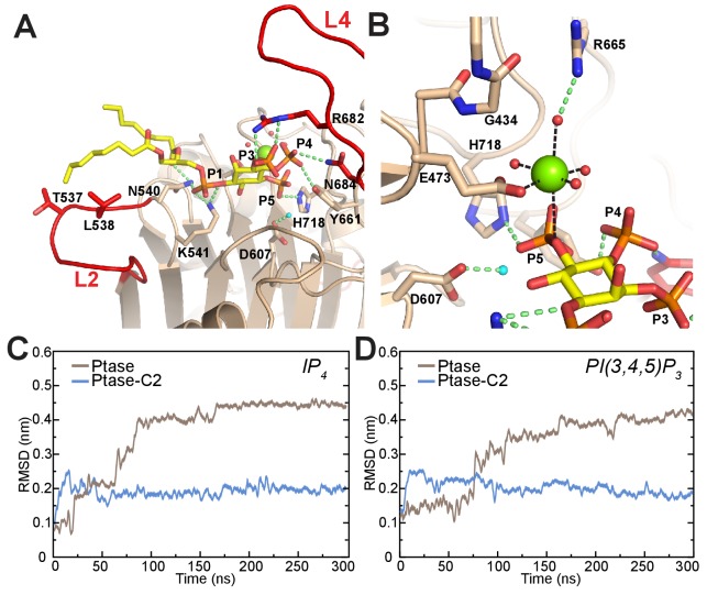
Figure 6. Model of substrate bound SHIP2.

(A) The SHIP2 Ptase-PI(3,4,5)P3-diC8 complex is modeled based on crystal structures of the homologue INPP5B crystal structure bound to PI(4)P (pdbs: 3MTC). L4 is in the ‘in’ conformation and R682 makes hydrogen bonds to the PI(3,4,5)P3 3-phosphate (P3). N540 and K541 interact with P1, N684 and Y661 with P4, H718 with P5 and L2 with PI(3,4,5)P3 lipid chains. The attacking water, bound to D607, is colored light blue and the Mg2+ ion is shown as green sphere. (B) Close-up of the Mg2+ coordination. (C–D) Plotted are RMSDs of the substrate headgroup atoms compared to the starting positions during MD simulations of the SHIP2 Ptase or Ptase-C2 bound to IP4 (C) or PI(3,4,5)P3-diC8 (D). The simulations are started with substrate positions according to the model shown in panel (A).

DOI: http://dx.doi.org/10.7554/eLife.26640.020

Figure 6.

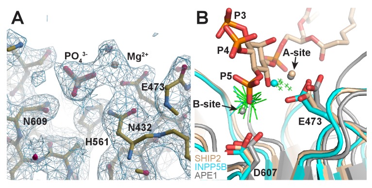
Figure 6—figure supplement 1. Structure of Ptase-C2 D607A, crystallized in presence of PI(3,4,5)P3-diC8 and Mg2+.

Figure 6—figure supplement 1.

(A) Electron density for one Mg2+ and one phosphate group is clearly visible in 6 of the eight molecules (shown is molecule H). 2Fo-Fc electron density is shown in blue countered at a σ level of 1.0. (B) Superposition of our model of the SHIP2 Ptase bound to PI(3,4,5)P3 and Mg2+ in the Michaelis-Menten complex (tan) with the 6 Ptase-C2 D607A molecules bound to Mg2+ and phosphate (green; only Mg2+ as small crosses and phosphates are shown), the Mg2+ and phosphate bound INPP5B structure (cyan, pdb: 5A7I) and the two-metal bound APE1 structure (grey, pdb: 1E9N). The E473 and D607 side chains labeling corresponds to SHIP2 numbering and the A- and B-metal sites in the APE1 structure are indicated.