
Figure 2. LPC-induced MAPK activation mediates p38 activation and subsequent STAT1 Ser727 phosphorylation.
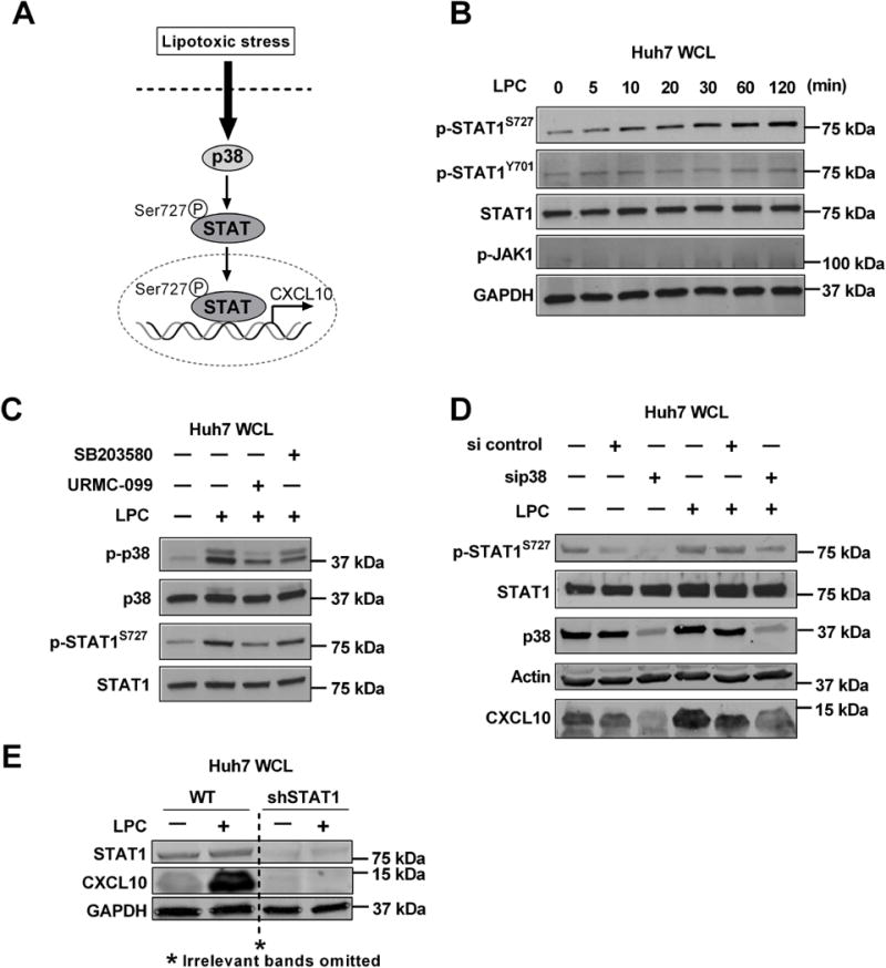
(A) Schematic representation of LPC-induced stress response resulting in p38 MAPK activation and STAT1 Ser727 phosphorylation, nuclear localization and transcriptional activation with subsequent CXCL10 induction. Immunoblot was used to assess (B) Phospho-STAT1 Ser727 and Tyr701, total STAT1, Phospho-JAK1 and GAPDH levels in whole cell lysates from Huh7 treated with 40 μM LPC at the time points indicated; (C) Phospho-p38 and Phospho-STAT1 Ser727and their respective total protein levels in whole cell lysates from Huh7 cells treated with either vehicle, or 40 μM LPC for 1 hr. with and without 1 μM URMC-099 or 10 μM SB203580; (D) Phospho-STAT1 Ser727, total STAT1, p38, actin and CXCL10 protein levels in whole cell lysates from Huh7 cells transfected with sip38 or scrambled siRNA and treated with vehicle or 40 μM LPC for 1 hr.; (E) STAT1, GAPDH, and CXCL10 protein levels in whole cell lysates from WT and shSTAT1 Huh7 cells treated with vehicle or 40 μM LPC for 1 hr.