
Figure 3. LPC-induced nuclear localization of phospho-STAT1 (Ser727) is MLK3/p38-dependent.
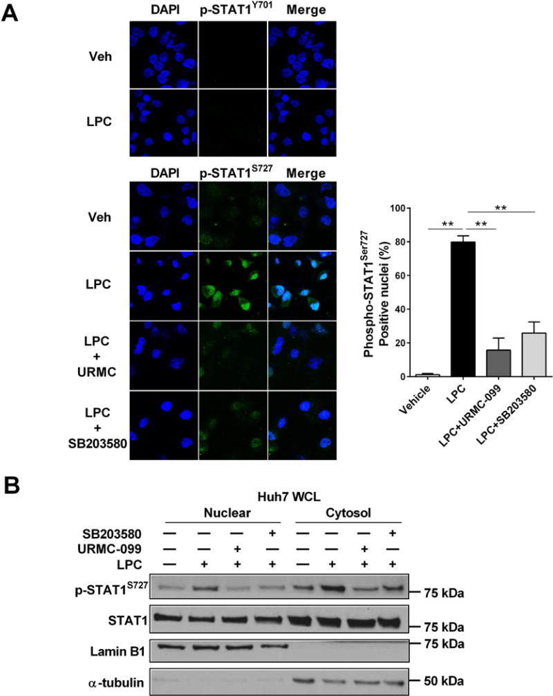
(A) Huh7 cells were treated with either vehicle or 40 μM LPC with or without 1 μM URMC-099 or 10 μM SB203580, a pharmacological p38 inhibitor, for 30 minutes. Nuclear localization of Phospho-Tyr707 and Phospho-Ser727 STAT1 were examined by immunocytochemistry and confocal microscopy. The Phospho-Ser727 STAT1 positive nuclei cells were quantified in five random 20 × microscopic fields. (B) Immunoblot analysis was used to assess Phospho-Ser727 STA1, STAT1, and GAPDH protein levels in the cytosolic fraction and Phospho-Ser727 STAT1, STAT1, and lamin B1 protein levels in the nuclear fraction from Huh7 cells treated with vehicle or LPC for 2 hours with or without 1 μM URMC-099 or 10 μM SB203580, a pharmacological p38 inhibitor. Bar columns represent mean ± standard error of the mean. ** p < 0.01