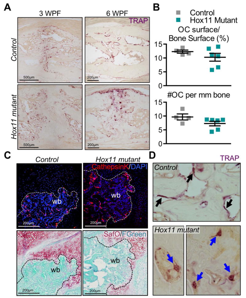
Figure 5. Osteoclasts are present, express markers of resorption in the Hox11 mutant callus.

(A) TRAP-stained callus sections from control and Hox11 mutant animals at 3 and 6 WPF show TRAP+ osteoclasts in calluses. (B) Histomorphometric quantification of osteoclasts per bone surface (%) and number of osteoclasts per 1mm of bone surface is comparable in controls and mutants. (C) Cathepsin K-stained callus sections from control and mutant animals show positive staining in controls and mutants. Safranin O/Fast Green staining on the same sections shows the overlap of CathepsinK with woven bone areas. (D) Images of large, detached osteoclasts in the Hox11 mutant callus compared to control osteoclasts that are flat and attached to the bone surface.
