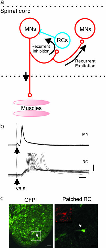
Fig. 1.
Examining MN synapses. (a) Schematic drawing of synapses formed by the MNs. (b) Paired intracellular recording of a MN and a RC. The MN is identified by its antidromic activation from ventral root stimulation (VR-S), and the RC is synaptically activated from VR-S at the same stimulus strength. (Scale bars: 20 mV and 5 ms.) (c) Transverse section of the lumbar spinal cord showing GFP-GAD67-positive cells; Left shows the enlargement of the RC region medial to the motor nucleus showing GFP-GAD67-positive cells (light green) and one GFP-GAD67-positive cell also filled intracellularly with Alexa Fluor 488 (bright green, arrow) that was also labeled with an antibody against calbindin (Inset). (Scale bars: Left, 50 μm; Right, 20 μm.)

