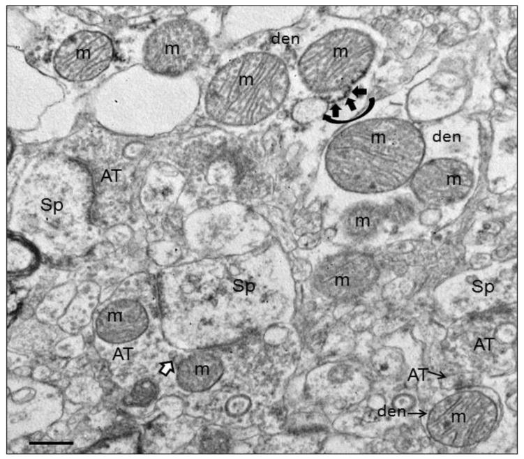
Figure 1.
Electron micrograph of human striatum. Mitochondria (m) are shown in various subcellular locations. In the dendrite (den) at the top of the field, mitochondrial associated ER (MAM) is shown (curved black arrow) with ER (short black arrows) connecting to the adjacent mitochondrion. In the terminal (AT) synapsing on a spine (Sp) in the lower part of the field, mitochondrial derived vesicles (MDVs) are shown (white arrow with black outline) budding off of a mitochondrion. Scale bars = 0.5 μm. Figure is modified from Figure 2a in Somerville et al., 2011b.

