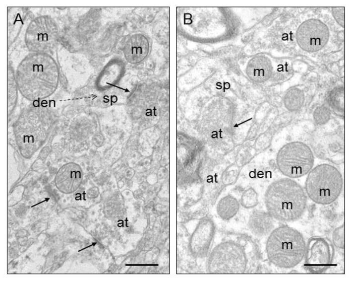
Figure 2.
Electron micrographs of human striatum. A) Control. B) Schizophrenia. Mitochondria (m) are shown in various subcellular locations. Abbreviations: s, spine; at, axon terminal; den, dendrite; ma, myelinated axon; solid arrows, synapses; dotted arrow, spine emerging from dendrite. Scale bars = 0.5 μm. Figure is modified from Figure 1 in Somerville et al., 2011b.

