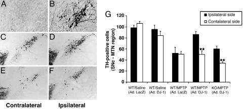
Fig. 5.
MPTP-induced neuronal loss is rescued by adenoviral expression of DJ-1. (A and B) DJ-1–/– mice were injected unilaterally in the SNc with adenoviral vectors expressing either DJ-1 or LacZ (control). Sections of the SNc at the medial terminal nucleus were immunostained with anti-DJ-1 antibody to detect adenoviral-mediated DJ-1 expression. (A) DJ-1 expression is not visible in the contralateral (uninjected) hemisphere of DJ-1–/– SNc. (B) Upon injection of vector expressing WT DJ-1, DJ-1 expression is restored to the ipsilateral hemisphere of DJ-1–/– SNc. (C–F) DJ-1+/+ and DJ-1–/– mice were injected unilaterally in the SNc with adenoviral vectors expressing either DJ-1 or LacZ (control; not shown). Injected animals were then treated with MPTP as for Fig. 2. SNc sections were prepared and immunostained as for A and B above. The contralateral hemisphere served as the noninjected, MPTP-treated control. (C)TH+ neurons are reduced on the contralateral (no DJ-1 injection) side of the SNc of an MPTP-treated DJ-1+/+ mouse when compared with (D) the ipsilateral (DJ-1-injected) side of the same mouse (E). Very few TH+ neurons are present in contralateral side of the SNc from an MPTP-treated DJ-1–/– mouse, reflecting the hypersensitivity of these animals to oxidative stress. (F) Overexpression of DJ-1 in the ipsilateral side of the DJ-1–/– SNc attenuates this neuronal loss. (G) Quantification of numbers of TH+ neurons in the contralateral and ipsilateral SNc hemispheres of all animals in the groups represented in A–F. Data in G are presented as mean ± SEM (n = 4–5 animals per group), where ** denotes a significance level (ANOVA) of P < 0.01.

