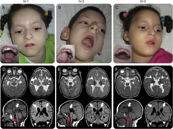
Figure 1. Clinical and neuroradiologic features of patients carrying homozygous AMP2 mutations.
(A–C) Facial images demonstrate shared features of microcephaly, sloping forehead, large and posterior rotated ears, and mandibular hypoplasia. In insets, mottled teeth with multiple cavities. (D–F) Brain MRIs from each patient showing characteristic “figure of 8” midbrain appearance (dotted ovals) and small hyperintense basal ganglia and thalami (arrowheads) on axial T2-weighted images. Sagittal T1-weighted images show small pons (red arrows) and extremely severe callosal hypoplasia (empty arrows). Coronal FLAIR images reveal hypoplasia/atrophy of the cerebellar hemispheres (arrowheads) with relative sparing of the vermis. Leukoencephalopathy similar to periventricular leukomalacia present in all patients, with loss of white matter bulk, periventricular hyperintensity, and enlarged lateral ventricles (empty arrows). FLAIR = fluid-attenuated inversion recovery.

