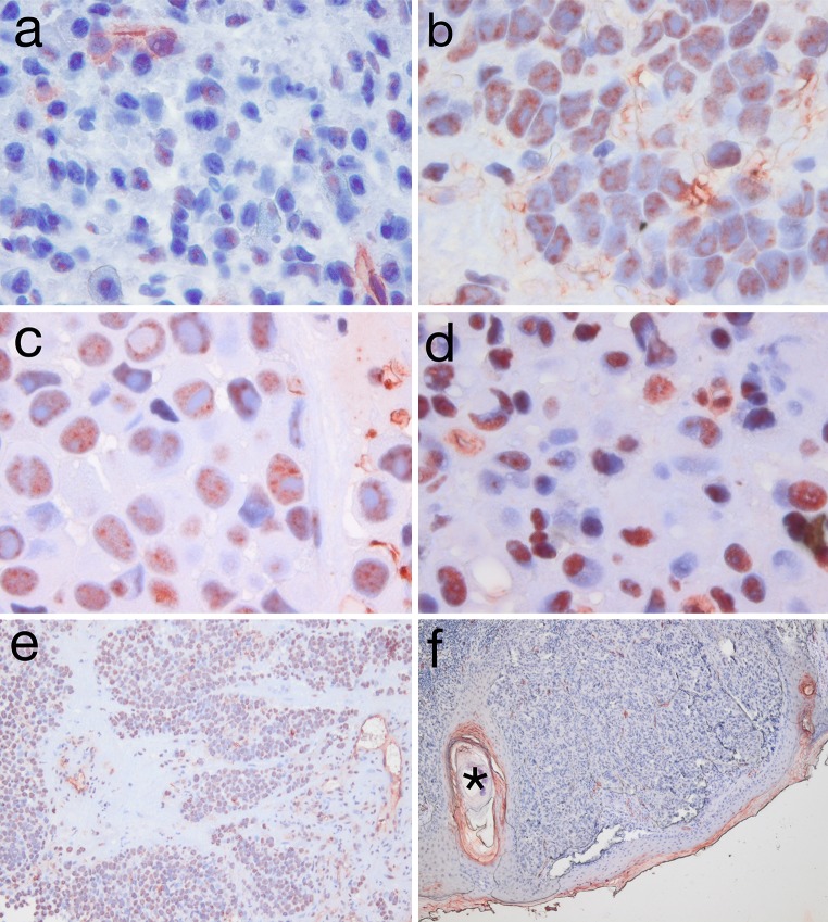
Fig. 1.
(×60 a–d; ×10 e + f): a + b: mucosal melanoma sample with score 1 (low; a) staining for KLK6 and score 3 (high; b) staining for KLK6; c + d: sample of mucosal melanoma with cytoplasmic (c) and cytoplasmic and nuclear staining for KLK6 (d); e + f: sample of mucosal melanoma with KLK6 in ×10 magnification (e), cutaneous melanoma without KLK6 expression but with internal positive control of a hair follicle with positive keratinocytes* (f); Since MMHN specimens showed a pigmentation in 50% of all cases, a bleaching method (7.5% H2O2 for 1 h) was used to lighten the brown pigmentation of the cells

