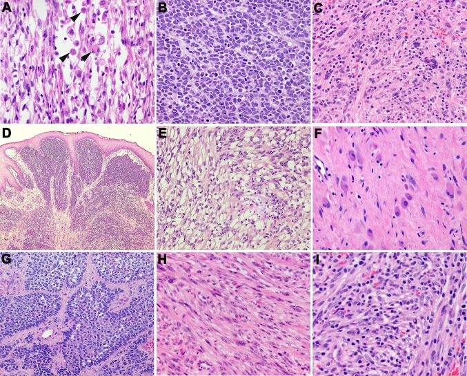
Fig. 2.
Histologic features of head and neck RMS. a Rhabdomyoblasts (indicated by arrowheads) in ERMS (Case 2); b ARMS with alveolar arrangement of tumor cells (Case 4); c PRMS with pleomorphic cell morphology including multi-nucleated cells (Case 6); d ERMS (Case 1) with sub-epithelial condensation of tumor cells, resembling a cambium layer; e Nests and clusters of ERMS cells with abundant clear cytoplasm (Case 2); f Prominent cytodifferentiation of ARMS cells into myoblast-like cells post-radiation (3); g Nests of ARMs with prominent palisading of tumor cells in the periphery (Case 5); h PRMS with areas of spindle cell morphology (Case 7); i PRMS with pleomorphic tumor cells with prominent lymphocytic infiltrate (Case 6)

