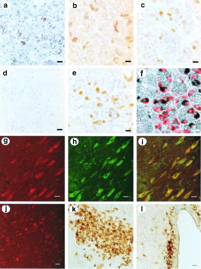
Figure 1.
Expression of type II GnRH receptor in pituitary and brain. Staining is shown in human (a), mouse (b), and sheep (c–f) pituitary. Sheep pituitary staining was neutralized by type II receptor peptide (d) but not by type I receptor peptide (e). The specificity of the staining was also indicated by the demonstration of similar staining by antisera raised in three other rabbits to the type II receptor peptide and an absence of staining by preimmune serum from all of these rabbits. Moreover, the hemocyanin carrier protein failed to neutralize staining. No staining was present with preimmune serum (not shown). Dual staining with type II receptor antiserum (black) and LH antiserum (red) shows colocalization (f). Type II GnRH receptor expressing neurones in the non-human primate brain by immunocytochemical staining is shown in panels g–l. Type II GnRH receptor-positive neurones are seen in the basal nucleus of Meynert (g), in the medial preoptic area (j) of an adult cynomologus monkey (Macaca fascicularis), and the amygdala (k) and periventricular region of the hypothalamus (l) of a fetal rhesus monkey (Macaca mulatta) at embryonic day 70. Note that the confocal photomicrographs with immunofluorescent staining show that the type II GnRH receptor-positive neurones (g, red-Rhodamine) in the basal nucleus of Meynert were also positive to mammalian GnRH I ligand (h, green-FITC). Colocalization is seen as yellow (i). In contrast, type II GnRH receptor-positive neurones in the medial preoptic area (j, red-Rhodamine), amygdala (k), and periventricular area (l) were negative for mammalian GnRH I ligand (data not shown). In k and l, immunoreactive products were visualized with DAB (brown). Scale bar: 10 μm for a–i; 20 μm for j–l. These brain areas also stained positive with antiserum raised against the carboxyl-terminal tail of the type II GnRH receptor (data not shown). Staining of type II GnRH receptor was neutralized in all tissues by incubation with type II peptide immunogen but not by type I peptide. Type II GnRH receptor-positive neurones were also seen in extrahypothalamic regions, such as medial septum, bed nucleus of the stria terminalis, substantia innominata, claustrum, amygdala, and putamen, and in the hypothalamic regions, such as supraoptic nucleus, ventromedial nucleus, and dorsomedial nucleus (data not shown).

