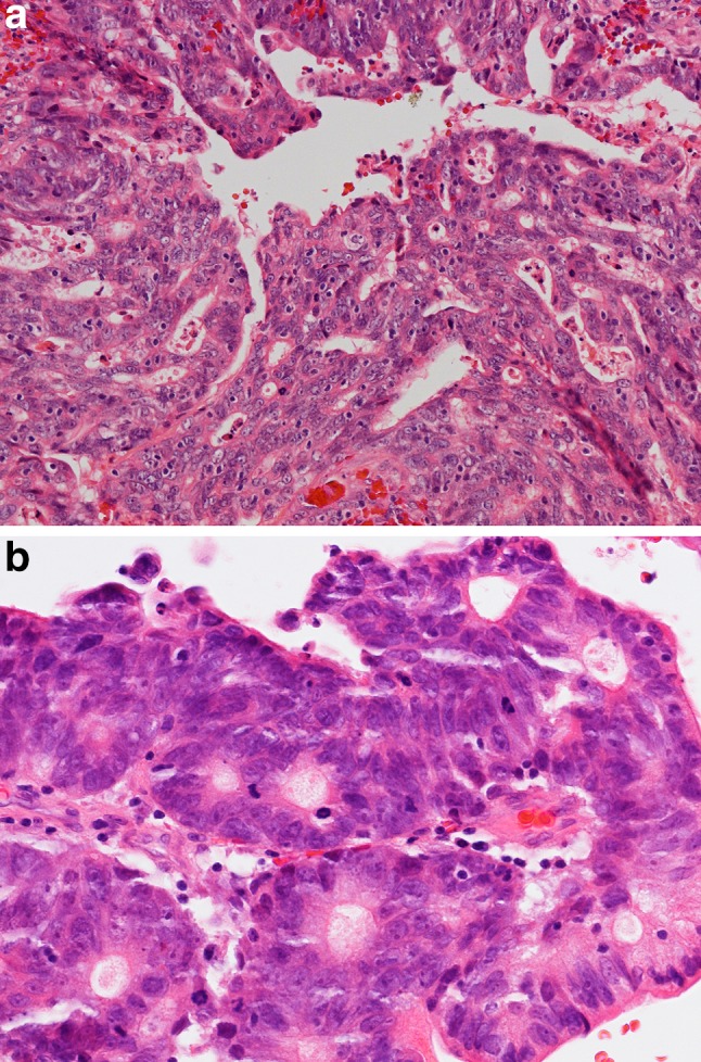
Fig. 2.

a Intestinal-type adenocarcinoma, colonic subtype. The tumor has glandular and trabecular areas resembling appearances of colorectal adenocarcinoma. H–E stain ×250. b The nuclei are crowded and highly pleomorphic and show some hyperchromasia. There is high mitotic activity. H–E stain ×400
