
Fig. 6.
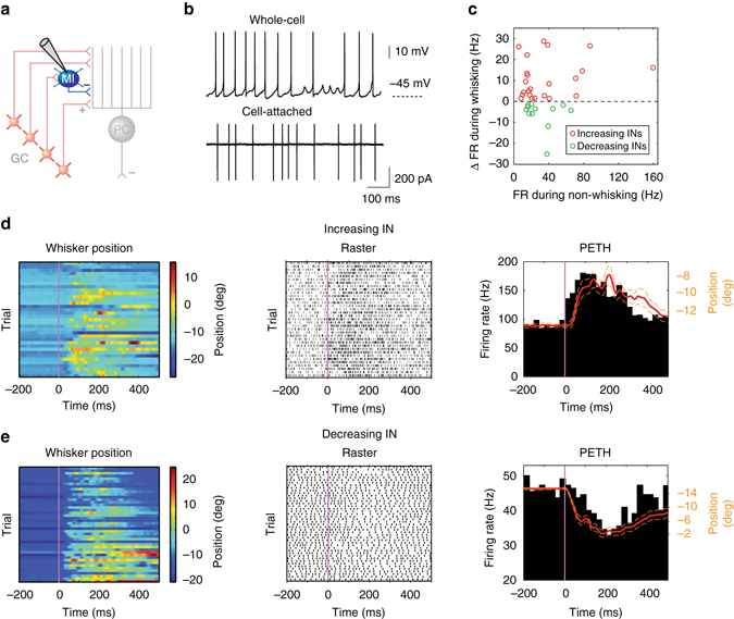
Firing rate alteration of inhibitory interneurons during whisking. a Schematic representation of cerebellar circuit highlighting inhibitory interneurons (INs) in the molecular layer. b Representative whole-cell and cell-attached recordings from INs. c Relative firing rate change during whisking with respect to baseline rate during non-whisking for all significantly modulated INs (P < 0.05, Wilcoxon signed-rank test, n = 35). Red and green circles represent increasing (n = 23) and decreasing (n = 12) cells, respectively. d IN exhibiting increasing firing rates during whisking. Colour-coded whisker movement (left) and the corresponding spike raster plot (middle). PETH from the same IN was overlaid with the mean whisker position (red; dashed orange lines: S.E.M) to show the close relationship between firing rate change and movement (right). e IN exhibiting decreasing firing rates during whisking. Colour-coded whisker movement (left) and the corresponding spike raster plot (middle). PETH from the same IN was overlaid with the mean whisker position (red; dashed orange lines: S.E.M) to show the close relationship between firing rate change and movement (right)