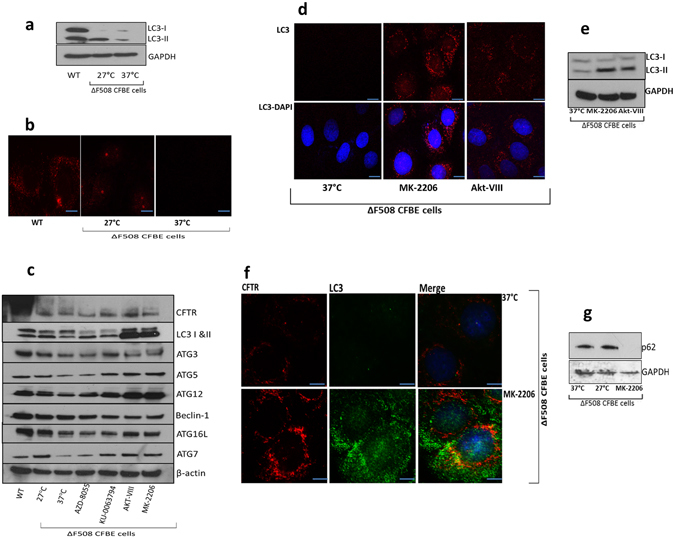
Figure 4.

Autophagy is restored in ΔF508 CFBE41o- cells upon treatment with MK-2206 and AKT-VIII. (a) Protein expression of LC3-I, LC3-II was determined by immunoblotting in WT HBE41o- cells, ΔF508 CFBE41o- cells (37 °C) and ΔF508 CFBE41o- cells temperature shifted to 27 °C. (b) Subcellular localization of LC3 (red- 549 nm) was detected in WT HBE41o- cells, ΔF508 CFBE41o- cells (37 °C) and CFBE41o- cells temperature shifted to 27 °C. (c) ΔF508 CFBE41o- cells (37 °C) were treated with AZD-8055, KU-0063794, AKT-VIII and MK-2206 and protein expression of autophagy markers was determined by immunoblotting (LC3-I and -II, ATG3, ATG5, ATG7, ATG12, ATG16l and beclin-1) in addition to CFTR and β-actin. WT HBE41o- cells and ΔF508 CFBE41o- cells at 27 °C were included as positive controls and ΔF508 CFBE41o- cells at 37 °C were included as negative controls. (d) Fluorescent detection was used to visualise LC3 (red- 549 nm) in ΔF508 CFBE41o- (37 °C) treated with MK-2206 and AKT-VIII (e) ΔF508 CFBE41o- cells (37 °C) were treated with AKT-VIII and MK-2206 along with negative control ΔF508 CFBE41o- cells at 37 °C and protein expression of LC3-I, LC3-II and GAPDH was determined by immunoblotting. (f) Co-localisation of CFTR (red- 549 nm) and LC3 (green- 488 nm) in ΔF508 CFBE41o- cells treated with MK-2206 and a negative control CFBE41o- cells at 37 °C. Nuclei were stained with DAPI (blue- 358 nm). Scale bar is 10 µm. (g) Protein expression of p62 was determined in ΔF508 CFBE41o- cells (37 °C), ΔF508 CFB41o-E cells (27 °C) and ΔF508 CFBE41o- cells treated with MK-2206.
