Figure 2.

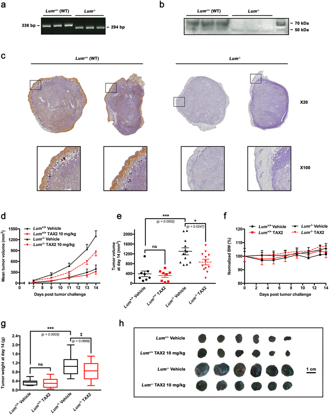
Evaluation of endogenous lumican impact on tumor growth and response to TAX2 treatment in a melanoma allograft model. (a) Genotype of representative offsprings from wild type (Lum +/+) matings and lumican deficient (Lum −/−) matings by PCR analysis. (b) Western immunoblotting on total protein extracted from skin of wild type (Lum +/+) mice (n = 3) and lumican-deficient (Lum −/−) mice (n = 3) with a rabbit polyclonal antibody raised against lumican core protein. Protein extracts from wild type mice exhibit a broad band at 57–90 kDa indicating the presence of glycosylated lumican as expected. Skin from lumican-deficient (Lum −/−) animals lacks any immunoreactive material confirming the absence of any lumican gene product. (c–h) B16F1 melanoma cells (2.5 × 10⁵) were s.c. inoculated in Lum +/+ or Lum −/− syngeneic C57BL/6 J mice, and then i.p. administrations of either Vehicle (0.9% NaCl) or TAX2 peptide (10 mg/kg) were performed on days 3, 5 and 7 post tumor cells inoculation. On day 7, tumors were detectable and tumor volume was measured every 1–2 days as described in Materials and Methods. (c) Microscopic views of s.c. allograft whole sections IHC (top panel, ×20) allowing visualization of lumican within tumors implanted in wild-type (Lum +/+) mice while it is neither detected in tumors from Lum −/− animals. Insets show higher magnification (×100) of stromal margin surrounding melanoma allografts. (d) Averages of calculated tumor volumes in mm3 (mean ± SEM, n = 8–12 per group). (e) Scatter dot plot of individual calculated tumor volumes on day 14. Line, mean ± SEM (t test, ns not significant, *p < 0.05, ***p < 0.001). (f) Evolution of normalized mice body weights (BW), expressed as a percentage of day 0 (mean ± SEM). (g) Tukey’s box and whisker plots of isolated tumor weight in g. Significant (***p < 0.001) as well as marginally significant (‡ p < 0.10) differences between groups are indicated (t test). (h) Representative photographs of B16F1 melanoma s.c. allografts after tumor excision.
