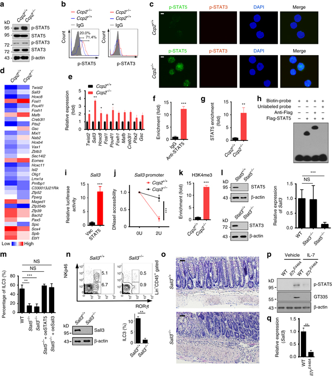
Fig. 5.

IL-7Rα glutamylation promotes Sall3 expression by STAT5. a–c Phosphorylation of STAT3 and STAT5 was tested by western blotting a, flow cytometry b and immunofluorescence staining c in CHILPs from WT and Ccp2 −/− mice after IL-7 stimulation. p-STAT5, green; p-STAT3, red; nucleus, blue. d Heat map of representative gene expression values from microarray data. In all, 1 × 10⁶ CHILPs (Lin−Flt3−CD25−IL-7Rα+α4β7 +) from Ccp2 +/+ or Ccp2 −/− mice were sorted for microarray. e Analysis of indicated gene expression levels in Ccp2 +/+ and Ccp2 −/− CHILPs by quantitative reverse transcription PCR (RT-qPCR). Relative fold changes of gene expression values were normalized to endogenous Actb. f, g Enrichment assessment of STAT5 on Sall3 promoter in CHILPs from Ccp2 +/+ and Ccp2 −/− mice. h The association of STAT5 with Sall3 promoter was examined by EMSA. Sall3 promoter probe was biotin-labeled. i Flag-STAT5, pTK, and pGL3- Sall3 promoter were transfected into 293T cells for luciferase assay. j DNaseI accessibility of Sall3 promoter in CHILPs from Ccp2 +/+ and Ccp2 −/− mice was assessed. k H3K4me3 enrichment on Sall3 promoter was determined. CHILPs were isolated from Ccp2 +/+ and Ccp2 −/− mice, followed by ChIP assay. l Sall3 mRNA levels were detected in WT, Stat3 −/− , and Stat5 −/− CHILPs. m CHILPs were isolated from WT mice and cultured with mitomycin C-treated OP9 cells for in vitro differentiation assay. Stat5 −/− CHILPs were transfected with STAT5 or Sall3 overexpression plasmid. Percentages of ILC3s were analyzed by flow cytometry. oe overexpression. n ILC3s were analyzed in Sall3 +/+ and Sall3 −/− mice. n = 6 for each group. o Histology of colons from Sall3 +/+ and Sall3 −/− mice 8 days after C. rodentium infection. Scale bars, 50 μm. p IL-7Rα glutamylation and STAT5 phosphorylation in CHILPs in the absence or presence of IL-7. CHILPs from WT or Il7r E446A BM were incubated with IL-7 and vehicle, followed by immunoblotting. β-actin was probed as loading controls. q Sall3 expression levels were tested in WT and Il7r E446A CHILPs by RT-qPCR. Relative fold change of Sall3 expression values were normalized to endogenous Actb. **P < 0.01, ***P < 0.001 (Student’s t-test). Data represent three independent experiments. Error bars in e–g, i–n, and q indicate s.d.
