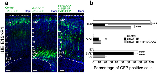
Figure 3.

Activation of phosphatidyl inositol-3 kinase (PI3K) rescues migration defects. (a) Coronal sections of P4 brains electroporated at E15 with shIGF-1R/CAG-GFP, control shRNA/CAG-GFP or shIGF-1R/CAG-GFP together with a constitutively active form of PI3K (p110CAAX-right); Calibration bar = 100 μm. (b) Quantification of the distribution of GFP-positive cells in VZ/SVZ/IZ, and layers II-IV and V-VI as indicated in (a). Note the significant increase in GFP positive cells in the deep layers (VZ/SVZ/IZ) zones and the decrease of GFP positive cells in the upper layers (II-IV) when knocking down IGF-1R. Co-transfection with p110CAAX rescued migration defects Post hoc Turkey’s ANOVA **p ≤ 0.01, ***p ≤ 0,0001. n = 3 independent experiments. An average of 500 cells was scored for each condition.
