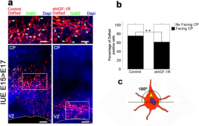
Figure 5.

shRNA-IGF-1R disrupts the polarized location of migrating neurons. (a) Co-electroporation of shIGF-1R/DsRed and GalT2-YFP into brains at E15 (left) decreases the proportion of DsRed-positive cells in the VZ and/or IZ with a Golgi apparatus oriented towards the radial axe fate at E17 compared to a control vector (left). Lower magnification pictures (bottom) and higher magnification insets (top) are shown. Calibration bar = 50 μm (bottom) or 25 μm (top). (b) Quantification of the experiment shown in A. Student’s t test; **p-value = 0.01. Note that in the brains suppressed for IGF-1R the number of cells facing the cortical plate is close to 60% (random distribution = 50%). n = 3 independent experiments. An average of 300 cells was scored for each condition. (c) For quantifications, we considered that a cell has the Golgi not oriented to the CP when the majority of the GalT2 staining was concentrated below the axis.
