
Figure 7.
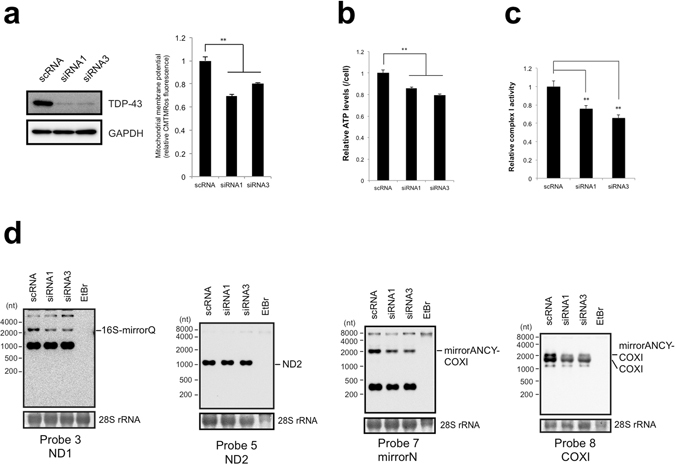
TDP-43 deficiency impairs mitochondrial function and decreases intermediates of mitochondrial transcripts. (a) Mitochondrial membrane potential was measured by CMTMRos staining of MCF7 cells after knockdown of TDP-43 with siRNA1 or siRNA3. The membrane potential per concentration of total proteins of the siRNA-treated cells relative to that for scRNA-treated cells harvested at 72 h after treatment is shown. Values are the average ± SD of five independent experiments. **P < 0.01 (unpaired t test). TDP-43 level was examined by western blotting with the indicated antibodies. (b) Cellular ATP levels per siRNA-treated MCF7 cell were calculated relative to that for scRNA-treated cells. Values are the average ± SD of six independent experiments. **P < 0.01 (unpaired t test). (c) Activity of electron transfer complex I per concentration of total proteins was measured for MCF7 cells harvested at 72 h after siRNA treatment relative to that measured for MCF7 cells with scRNA treatment. Values are the average ± SD of three independent experiments. **P < 0.01. (d) mt-tRNA levels upon knockdown of TDP-43 in MCF7 cells were assessed with northern blotting. Probes are indicated under the figures. The intermediates of mitochondrial transcripts were indicated to the right side of the figures. 28S rRNA stained by methylene blue served as a loading controls.