
Figure 5.
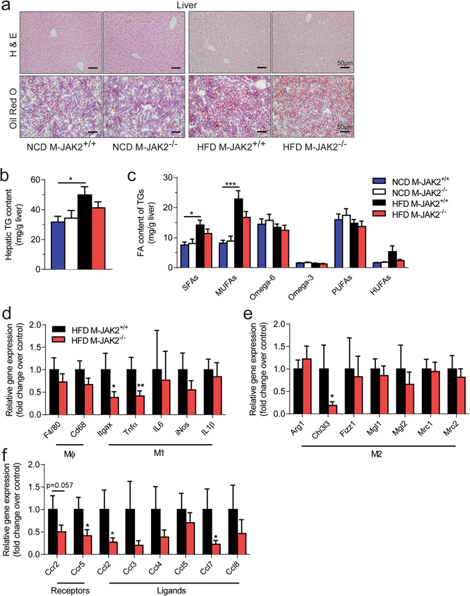
Reduced hepatic inflammation in HFD fed M-JAK2−/− mice. (a) Representative micrographs of H&E (top) and Oil Red O (bottom) stained liver sections from NCD or HFD fed M-JAK2+/+ and M-JAK2−/− mice. Quantification of (b) total triacylglycerol (TG) levels and (c) amount of saturated (SFAs), mono-unsaturated (MUFAs), Omega-6, Omega-3, polyunsaturated (PUFAs) and highly unsaturated (HUFAs) fatty acids (FA) of TGs in liver from NCD- (n = 4–6) and HFD- (n = 9–10) fed M-JAK2+/+ and M-JAK2−/− male mice. mRNA expression in liver from HFD-fed M-JAK2+/+ and M-JAK2−/− mice for (d) macrophage (Mϕ) and M1 markers, (e) M2 markers, and (f) chemokine receptors and its associated ligands (n = 8–9). All results are mean ± SEM; *p < 0.05, **p < 0.01, ***p < 0.001.