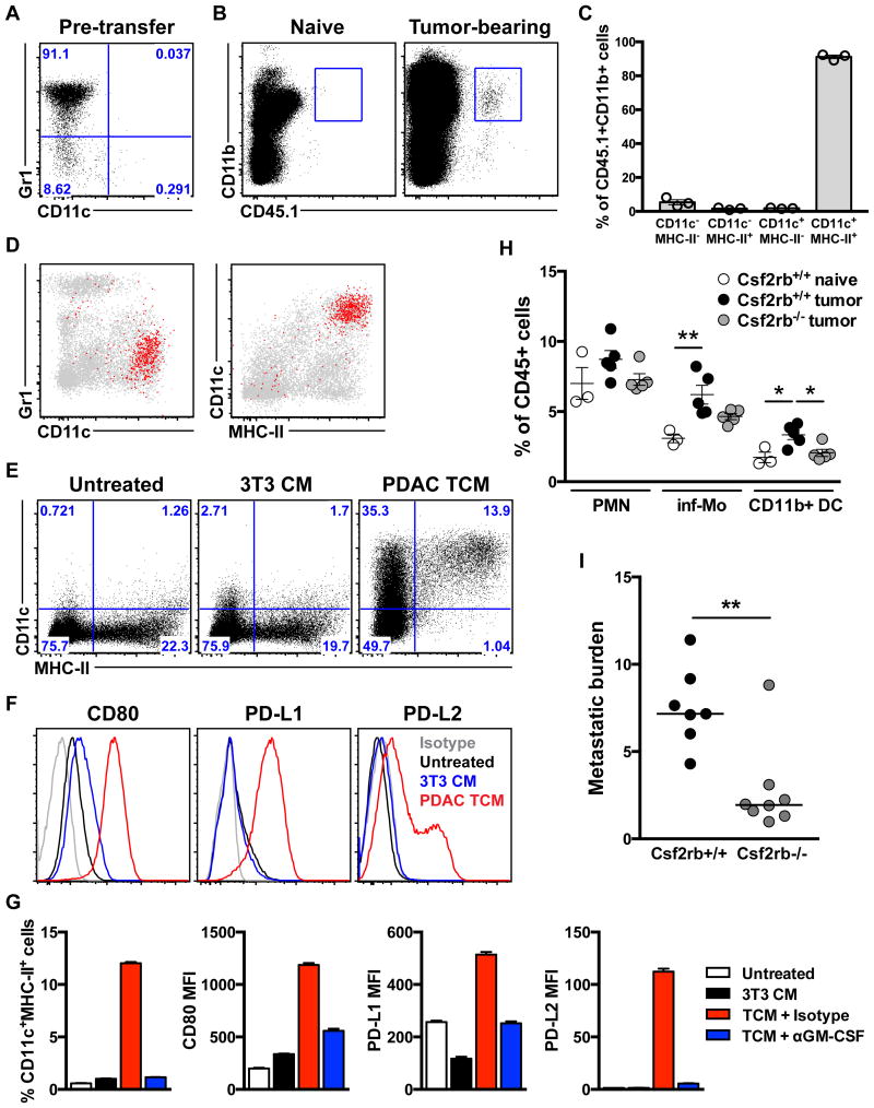
Figure 3. Metastasis-associated CD11b+ DC develop from monocytes in response to tumor-released GM-CSF.
(A-D) BM Mo from naïve CD45.1×129 F1 mice were transferred into naïve or 3.5 wk tumor-bearing mice and analyzed by flow cytometry after 5 d. (A) Phenotype of BM Mo prior to transfer. (B) Plots depicting recovery of donor-derived cells from the livers of tumor-bearing but not naïve mice. (C, D) Phenotype of CD45.1+CD11b+ cells (red) overlaid on host hematopoietic cells (gray) (D) and percentages of donor cells displaying the indicated phenotype with means ± SEM shown (C). (E-G) BM Mo from naïve mice were cultured for 18-24 hr in the presence of control or conditioned media ± the indicated antibodies, and then total CD11b+ cells were analyzed for the expression of the indicated markers. (H, I) BM chimeras were generated using WT (Csf2rb+/+) or GM-CSFR KO (Csf2rb-/-) donor cells and used in the experimental liver metastasis model. (H) CD11b+ subset frequencies in the liver 12 d after sham operation or tumor cell injection with means ± SEM shown. (I) Metastatic burden 21 d following tumor cell injection with medians shown. *, p<0.05; **, p<0.01 by one-way ANOVA with post hoc Tukey's test (H) or Mann-Whitney U-test (I). See also Fig. S2.

