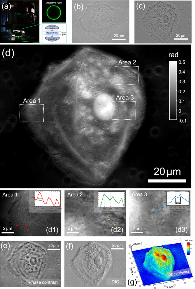
Figure 12.

High-resolution imaging of buccal epithelial cell. (a) Optical configuration with the corresponding rear focal plane image of the objective. (b) In-focus intensity image. (c) Intensity difference image. (d) Reconstructed phase image. Three areas of interest (boxed regions) are enlarged in (d1–d3). The insets shows line profiles taken at different positions in the cell. (e) Simulated phase-contrast image; (f) Simulated DIC image; (g) Pseudo-color 3D rendering of the cell optical length.
