Fig. 1.
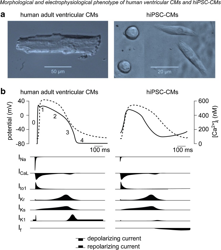
Morphological and electrophysiological phenotype of human-induced pluripotent stem cell-derived cardiomyocytes (hiPSC-CMs) and native human ventricular cardiomyocytes (CMs). a Morphological differences between human adult ventricular CMs and hiPSC-CMs; note the different scale. b Examples of action potential (solid line) and calcium transient (dashed line) in human adult ventricular CMs and hiPSC-CMs (upper panels), and schematic representation of the time course and magnitude of relevant ion currents (lower panels). Figure modified from [68] (a) and [15] (b)

