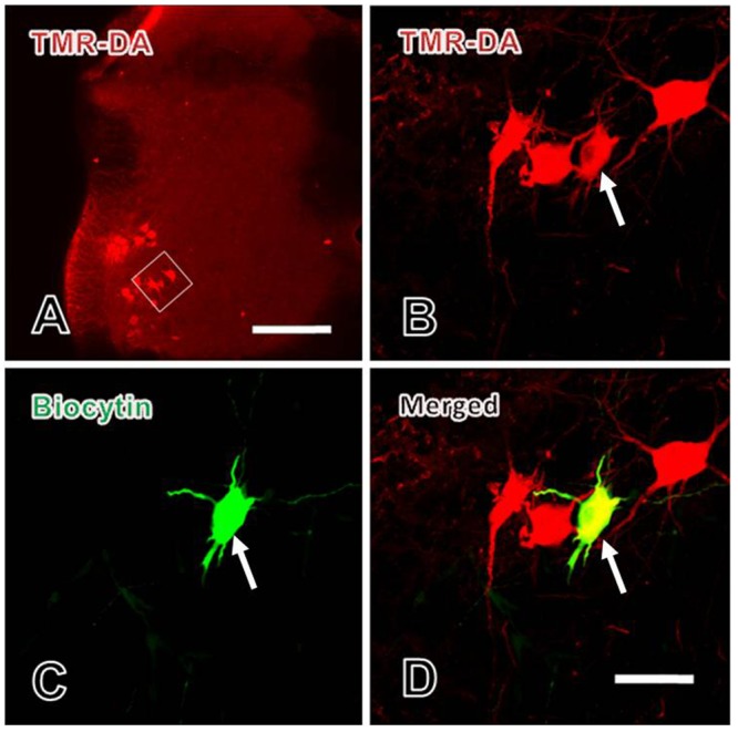
FIGURE 4.

Verification of the patch-clamp recorded neurons in spinal ventral horn. (A) The lower power image of TMR-DA labeled motoneuron (red) in lamina IX of spinal ventral horn in which TMR-DA was injected into the sciatic nerve. The white rectangle in (A) was magnified in (B). (B–D) The higher power images of the TMR-DA labeled motoneuron (red in B) also showed biocytin positive labeling (green in C), i.e., the TMR-DA/biocytin double labeled motoneuron (yellow in D, indicated with white arrows) in lamina IX of the spinal ventral horn. Scale bars = 200 μm in (A), 25 μm in (B–D).
