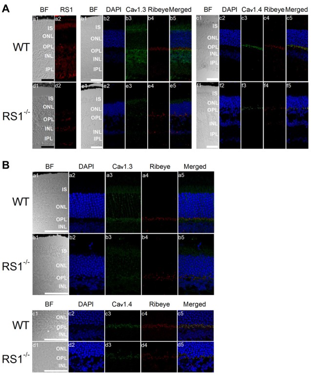
Figure 4.

Deletion of RS1 decreases the protein expression of Cav1.3 and Cav1.4. (A) The upper panel (a1-c5) contains retinal sections of wild type (WT), and the lower panel (d1-f5) contains retinal sections of RS1−/−. (a1-a2) and (d1-d2) are the immunostaining for RS1. (b1-b5) and (e1-e5) are the double immunostaining for Cav1.3 and Ribeye; (c1-c5) and (f1-f5) are the double immunostaining for Cav1.4 and Ribeye. The scale bar represents 50 μm. (B) The same immunostained retinal sections are shown at a higher magnification (40×). The upper panel contains retinal sections from WT (a1-a5) and RS1−/− (b1-b5) that were double-stained for Cav1.3 and Ribeye. Images in (a1-a5) and (b1-b5) include retinal layers of IS, ONL, OPL and INL. The lower panel contains retinal sections from WT (c1-c5) and RS1−/− (d1-d5) that were double-stained for Cav1.4 and Ribeye. Images in (c1-c5) and (d1-d5) include retinal layers of ONL, OPL and INL. The scale bar represents 50 μm. 4′s,6-diamidino-2-phenylindole (DAPI) stains the nuclei. BF, bright field; IS, photoreceptor inner segments; ONL, outer nuclear layer; OPL, outer plexiform layer; INL, inner nuclear layer; IPL, inner plexiform layer.
