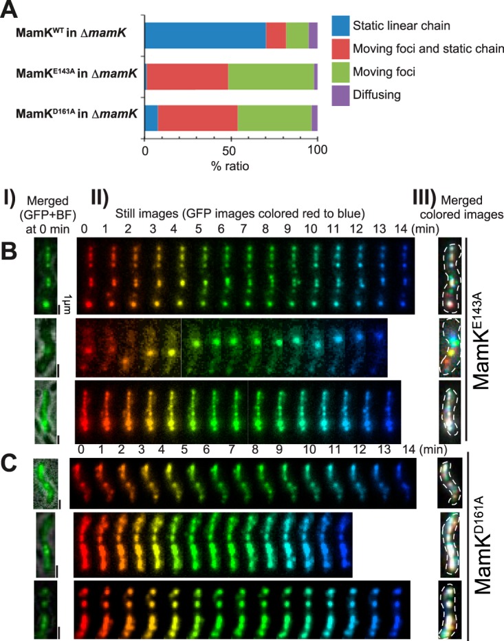
FIG 4 .

MamK ATPase activity is required for anchoring magnetosomes. (A) Dynamic localization patterns of magnetosomes. The dynamics of magnetosomes were determined using time-lapse imaging for 3 h in ΔmamK mutant cells expressing wild-type (WT) MamK (n = 192), MamKE143A (n = 151), or MamKD161A (n = 380), respectively, with MamC-GFP. (B and C) Effects of expression of ATPase-defective MamK, (B) MamKE143A, and (C) MamKD161A on the dynamics of magnetosomes. (Column I) Merged GFP and bright-field images of the cells at time zero. (Column II) Time-lapse images acquired for 14 min at 1-min intervals and sequentially rainbow colored from red to blue. (Column III) Merged images of the rainbow-colored still images in (II). Note that a single cell expressing MamKE143A and MamKD161A contains static (white) and dynamic (colored) magnetosomes. Moreover, the dynamic magnetosomes continuously attached and detached to the long axis of the cell, suggesting that magnetosomes were loosely attached to the ATPase-defective MamK filaments.
