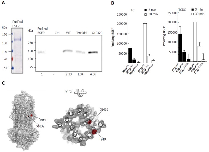
Figure 3.

In vitro transport assay of wild-type bile salt export pump, BSEP-T919del and BSEP-G1032R. A: Left, Coomassie brilliant blue (CBB) stained SDS-PAGE gel of purified Bile Salt Export Pump (BSEP) expressed in Pichia pastoris. 9 μg protein was loaded on the lane. Right, Western blot of purified BSEP and membrane preparations of BSEP-T919del, BSEP-G1032R, BSEP wild-type (WT) and empty control (Ctrl). 38.5 ng of purified BSEP or 15 μL of the membrane preparations were subjected to SDS-PAGE. After detection with the monoclonal antibody, the gel was analyzed densitometrically to calculate the amount of wild-type or mutant BSEP or in the membrane preparations (relative densitometric values below panel); B: [3H]-taurocholate (TC) and [3H]-taurochenodeoxycholate (TCDC) transport by wild-type BSEP, BSEP-T919del, and BSEP-G1032R after 5 (black bars) and 30min (white bars). Experimental details are provided in Materials and Methods. The amount of translocated TC (left) or TCDC (right) per mg of BSEP was determined based on the quantification of wild-type BSEP, BSEP-T919del and BSEP-G1032R in the membrane vesicle preparations. All measurements represent the average with standard deviation of triplicate measurements; C: The homology model of BSEP was generated based on the outward-facing crystal structure of the multidrug transporter Sav18662 from S. aureus (pdb entry: 2HYD). The putative localisation of T919 at the outer surface of the transporter molecule and of G1032 close to the potential translocation pore of BSEP is highlighted by red spheres.Pmol/mg BSEP. BSEP: Bile salt export pump.
