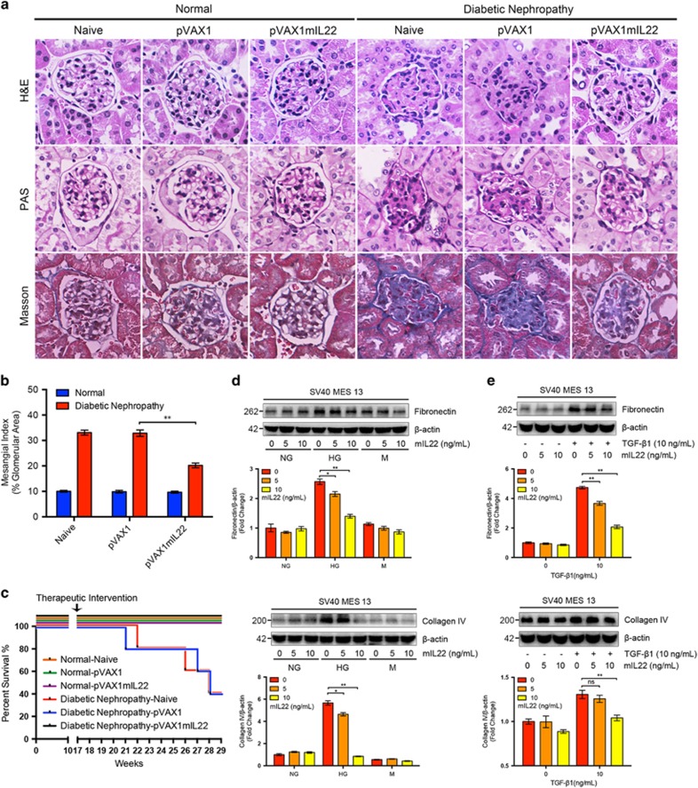
Figure 2.
Attenuated renal injury and mesangial matrix expansion with established diabetic nephropathy after IL-22 gene therapy. (a) Representative micrographs of H&E staining, PAS staining, and Masson staining. Original magnification: × 400. (b) Glomerular mesangial matrix expansion quantified from PAS staining. (c) Survival rate of mice after IL-22 gene therapy. (d–e) Reduced synthesis of high glucose-induced and TGF-β1-induced ECM proteins in renal glomerular mesangial cells after IL-22 treatment. Representative immunoblots and semi-quantitative analysis of cytosolic expression of fibronectin and collagen IV from three independent experiments was carried out using ImageJ (National Institutes of Health, Bethesda, MD, USA). Densitometric values of immunoreactive bands were normalized to those of β-actin and the results were expressed as fold changes. *P<0.05; **P<0.01; NS, no significance

