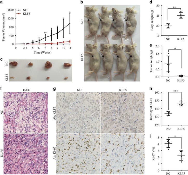
Figure 5.
KLF5 expression inhibits ccRCC xenograft growth in vivo. NC or KLF5-transduced 786-O cells were inoculated subcutaneously into nude mice. (a) Tumor volumes were measured every week and growth curves of two groups were shown. (b–e) Images of euthanized mice (b) and excised tumors (c) were shown. Bodyweight with tumor (d) and tumor weight alone (e) were measured. (f–i) Representative images of H&E staining (f) or IHC staining of KLF5 or Ki-67 (g) of these tumors were shown. Scale bar, 50 μm. Image J software was used to quantify the intensity of KLF5 (h) or Ki-67 positive cells (i) between NC and KLF5-transduced 786-O subcutaneous tumors. All bar graphs are plotted as mean±S.D. P-values are calculated between linked groups. *P<0.05; **P<0.01; ***P<0.001

