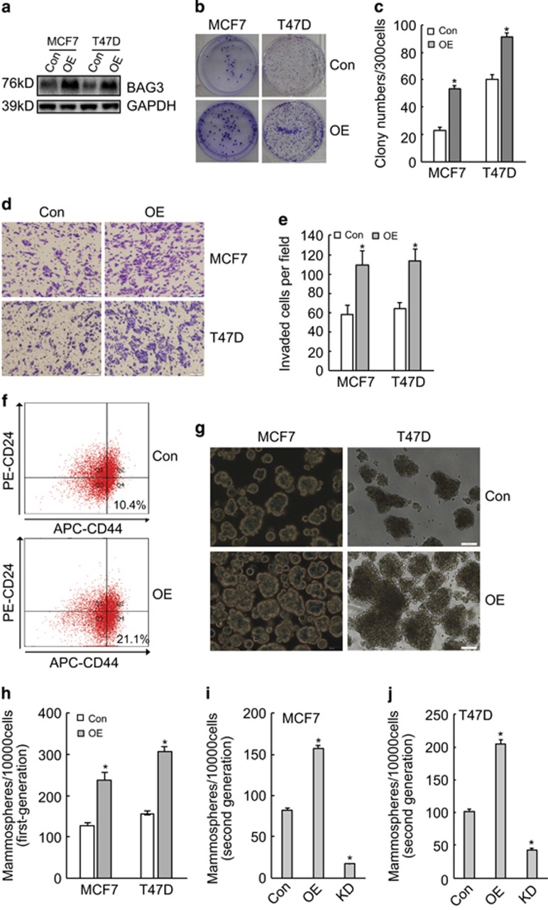
Figure 3.
Ectopic BAG3 promoted BCSC-like properties in vitro. (a) MCF7 and T47D cells were transduced with lentivirus containing empty (Con) or BAG3 construct (OE). BAG3 expression was analyzed using western blot. (b and c) Three hundred cells were plated on a six-well plate and cultured for 14 days. Representative photographs of plate colony formation were provided (b), and the number of colony was counted and represented as the mean±S.E.M. from three independent experiments (c). (d and e) The invasiveness of control or BAG3-overexpressing MCF7 and T47D cells was evaluated by a Matrigel-coated Transwell assay. Cells that passed through Matrigel for 24 h were stained with crystal violet. Representative photographs were provided (d), and cells were counted and represented as the mean±S.E.M. from three independent experiments (e). (f) The subpopulation of CD44+/CD24−/low cells from MCF7 cells was measured by flow cytometry. (g) Con or OE breast cancer cells were cultured under mammosphere-forming condition. Mammospheres were photographed by phase-contrast microscopy and representative images were provided. (h) The number of mammospheres was counted, and was represented as the mean±S.E.M. from three independent experiments. (i and j) Control, BAG3-overexpressng (OE) or BAG3 knockdown (KD) MCF7 (i) or T47D (j) cells were cultured under mammosphere-forming condition for 7 days; the first-generation mammospheres were disaggregated and single-cell suspensions were cultured under the traditional condition for 3 days, and then followed by floating culture. Mammospheres were photographed and the number of mammospheres was counted. NS, not significant; *P<0.01

