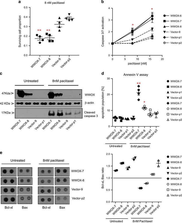
Figure 1.
WWOX-transfected PEO1 lines show increased apoptosis following treatment with paclitaxel, compared with vector-transfected lines. (a) Relative cell survival following treatment with paclitaxel for 72 h, measured by SRB assay. Means of quadruplicate experiments±S.E.M. (b) Caspase-3/7 activation following treatment with paclitaxel for 24 h, measured by CaspaseGlo3/7 reagent (Promega). Means of six experiments±S.E.M. (c) Immunoblot showing activated caspase-3 levels (MAB835 antibody; R&D Systems) following treatment with 8 nM of paclitaxel for 24 h. β-Actin bands (AC15 antibody; Sigma) show equal loading of blot and WWOX immunoblot (antibody gifted by Dr. M Aldaz, MD Anderson Cancer Centre) demonstrates successful knockdown. (d) Early apoptotic (Annexin V-positive/propidium iodide (PI)-negative) cells following treatment with paclitaxel for 24 h. Means of quadruplicate experiments±S.E.M. (e) Left panel: Bcl-xL and Bax probe spots from protein arrays, showing increased Bax and decreased Bcl-xL in WWOX-transfected lines following exposure with paclitaxel for 24 h; right panel: densitometry from protein array shows lower Bcl-xL:Bax ratio in WWOX-transfected cells treated with paclitaxel. Means of two probe spots±S.D. (*P<0.05; **P<0.01)

