Figure 1.
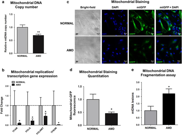
AMD cybrids have dysfunctional mitochondria. (a) AMD cybrids showed significantly reduced mitochondrial DNA (mtDNA) copy number compared to the age-matched normal cybrids (n=5, P=0.007). (b) AMD cybrids had lower expression levels of mitochondria replication and transcription genes. The AMD cybrids showed reduced expression of TFAM (90.1% decrease, P=0.04, n=5), POLG (78.1% decrease, P=0.03, n=5), POLRMT (53.8% decrease, P=0.03, n=5), and TFB2M (97.6% decrease, P=0.03, n=5) compared to normal cybrids (Supplementary Table S5). (c) Representative images of confocal microscopy showing diminished mtGFP staining throughout the cytoplasm in AMD cybrids compared to normal cybrids (Scale bar =20 μm). (d) Quantitation of the 1C images showed that AMD cybrids had a 54% (P=0.04, n=4) decrease in mtGFP fluorescence compared to normal cybrids, suggesting lower numbers of AMD mitochondria. (e) AMD cybrids showed higher number of mtDNA lesions within the 503–2484 bps region compared to normal cybrids (P=0.04, n=5). Data are represented as mean±S.E.M., normalized to normal cybrids (assigned value of 1). The endpoint for all experiments was 72 h

