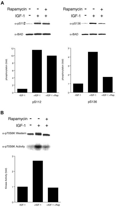
Figure 3.
Rapamycin specifically inhibits IGF-1-induced BAD S136 phosphorylation and p70S6K activity in the mitochondrial fraction. (A) Survival factors were withdrawn from FL5.12 BCL-XL/BAD S112A or FL5.12 BCL-XL/BAD S136A cells for 4 h (−IGF-1), followed by addition of IGF-1 (50 ng/ml) for 15 min (+IGF-1). Rapamycin (20 ng/ml) was added when factors were withdrawn. Western blot analyses were done with antibodies that recognize phosphorylated forms of BAD at S112 or S136 (19, 22). The filters were reprobed with an antibody that recognizes BAD regardless of its phosphorylation state. Data shown are representative of two independent experiments. Histograms present the fold increase in intensity of the phosphorylated band relative to total BAD, in which − IGF-1 is set at 1.0. (B) FL5.12 BCL-XL/BAD S112A cells were treated as in A. The mitochondrial heavy membrane fractions were analyzed by Western blot with an anti-p70S6K Ab (Upper), or immunoprecipitated with an anti-p70S6K Ab, followed by an in vitro kinase assay using GST-S6 as substrate. Data shown are representative of two independent experiments. Histogram presents the fold increase in intensity of the phosphorylated S6.

