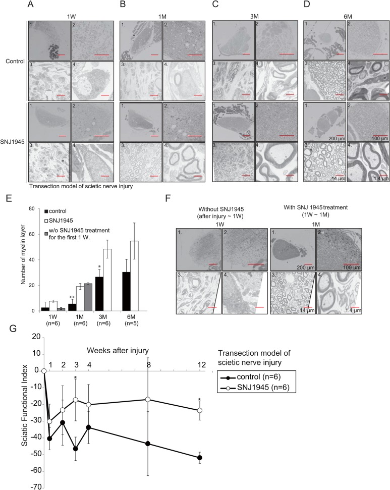
Fig. 7.
Facilitation of sciatic nerve regeneration by SNJ1945. Transection model of mouse SN injury. Imaging modality, orientation, and magnification are as follows: panel 1, low-magnification Prussian Blue staining; panel 2, high-magnification Prussian Blue staining; panel 3, electron microscopic (EM) image at low magnification; panel 4, EM at higher-magnification. Effects of oral SNJ1945 treatment on SN axon remyelination one week (A), one month (B), three months (C), and six months (D) after transection. Upper panels are from the untreated transection group (control). Lower panels are from mice treated with oral SNJ1945 after SN transection. SNJ1945 treatment accelerated remyelination of the sciatic nerve. (E) Statistical summary (mean±s.e. for 10 mice) showing enhanced numbers of regenerated myelinated SN axons in SNJ1945-treated mice after transection. *P<0.05 and **P<0.01 by ANOVA. (F) Effect of SNJ1945 on regeneration/remyelination when administration was delayed for one week after transection. Left panels: one week after transection (without treatment). Right panels: after one month of treatment. SNJ1945 treatment was still effective. (G) Gait analysis during recovery from SN transection. Sciatic functional index (SFI) of gait analysis indicating facilitated functional recovery after transection in SNJ1945-treated mice. *P<0.05 by Mann–Whitney U test. Error bars indicate standard errors.

