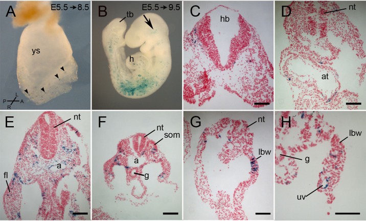
Fig. 3.
Lineage tracing of a cohort of cells that expressed Tbx6-creERT2 between E6.5 and E7.0. Pregnant Cre-reporter females were injected with tamoxifen at E5.5 and embryos were dissected at E8.5 (A) or E9.5 (B-H) and stained for lacZ. (A) An E8.5 unturned, whole mount embryos with scattered lacZ-positive cells (arrowheads) in the headfolds, lateral mesoderm, primitive streak and base of the allantois. (B) A whole mount E9.5 embryo of approximately 15-20 somites with lacZ-positive cells in the head (arrow), heart, and lateral mesoderm including somites, with lower concentrations in the rostral and caudal somites. (C-H) Representative transverse sections of E9.5 embryos showing lacZ-positive cells in the head mesenchyme at the level of the hindbrain (C), in the atrium of the heart (D), in the endothelium of the dorsal aorta and umbilical vein (E,H), somites, lateral body wall, forelimb bud, intermediate mesoderm and splanchnopleure mesoderm. a, aorta; at, atrium; fl, forelimb bud; g, gut; h, heart; hb, hindbrain; hf, headfold; lbw, lateral body wall; nt, neural tube; som, somite; tb, tail bud; uv, umbilical vein; ys, yolk sac. Compass in A refers only to panel A. Scale bars: 100 μm.

