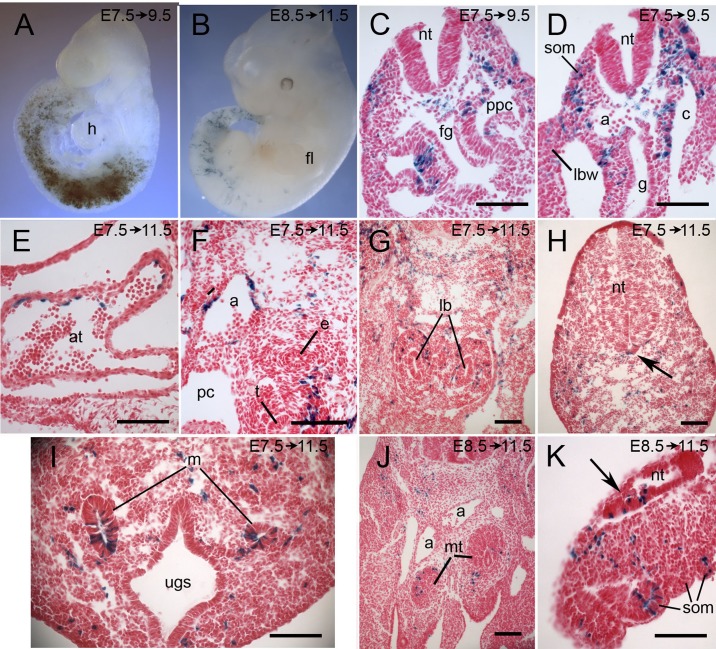
Fig. 5.
Lineage tracing of cohorts of cells that expressed Tbx6-creERT2 between E7.75 and E9 or between E8.75 and E10. Pregnant Cre-reporter females were injected with tamoxifen at E7.5 (A,C-I) or E8.5 (J,K) and embryos were dissected 2-4 days later as indicated on each panel and stained for lacZ. (A) A whole-mount embryo labeled at E7.5 with the bulk of the lacZ-positive cells in the somites and lateral mesoderm. (B) A whole-mount embryo labeled at E8.5 with a pronounced posterior shift in the distribution of labeled cells. (C-K) Transverse sections of embryos showing representative tissues with labeled cells, notably somites (C,D,K), lining of the coelom and pericardio-peritoneal canal (C,D), endothelium of the heart and aorta (D-F), mesenchyme surrounding the lung buds and trachea (F,G), mesonephric tubules and metanephric blastema (I,J), and tail mesenchyme (H,K). In a small number of embryos, labeled cells were seen in the posterior neural tube (arrow in H, in the floorplate of the tail neural tube; arrow in K, in tangential section of tail neural tube). a, dorsal aorta; at, atrium; c, coelom; e, esophagus; fg, foregut; fl, forelimb; g, gut; h, heart; lb, lung buds; lbw, lateral body wall; m, mesonephric duct; mt, metanephric blastemal; nt, neural tube; pc, pericardial cavity; ppc, pericardio-peritoneal canal; som, somite; t, trachea; ugc, urogenital sinus. Scale bars: 100 μm.

