
Fig. 5.
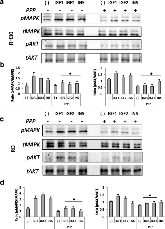
Effect of PPP treatment on INS, IGF1, and IGF2 signaling in RMS cells. RH30 (panel a) and RD (panel c) cells, untreated or PPP-treated (0.1 μM) for 72 h, were stimulated by INS (10 ng/mL), IGF1 (100 ng/ml), or IGF2 (100 ng/mL) for 5 min, and phosphorylation was assessed by western blotting. The experiment was repeated three times with similar results. Panels b and d show Western blot quantitation, expressed as a ratio of phosphorylated protein to total. *p < 0.05