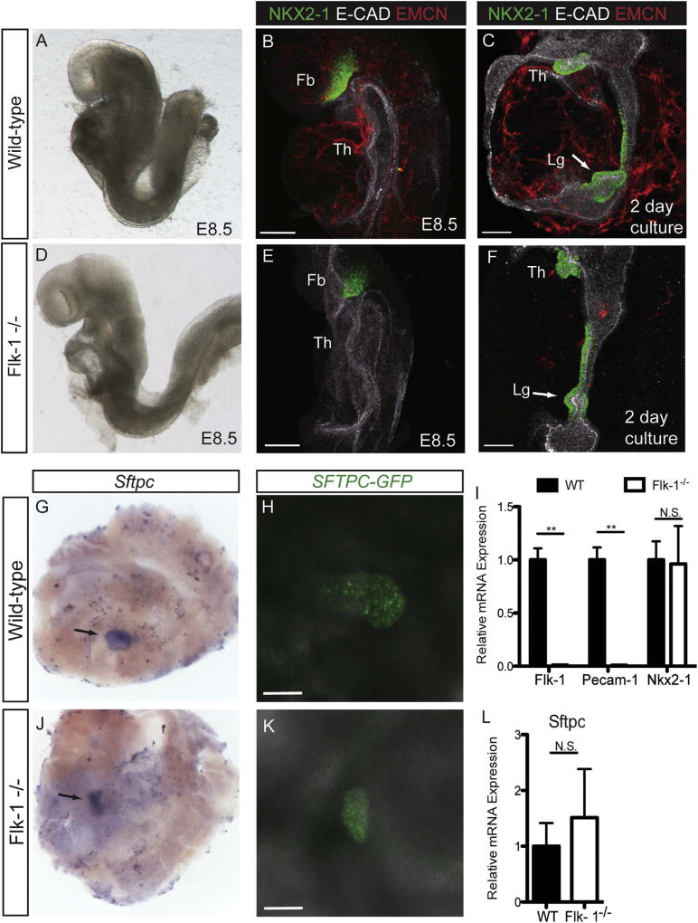
Fig. 4.
E8.5 Flk-1−/− mutants specify lung progenitors in vitro. Wild-type (A) and Flk-1−/− mutant (D) embryos are grossly indistinguishable at E8.5 (8–10ss). EMCN-positive endothelial cells are detectable in wild-type (B), but not Flk-1−/− mutant (E) embryos at E8.5. Immunofluorescent staining for NKX2-1 shows that after 2 days in culture, both wild-type (C) and Flk-1−/− (F) foreguts contain NKX2-1-positive progenitors in the lung field (arrows). qPCR analysis of Flk-1 and Pecam-1 expression in cultured explants (I) confirms the absence of endothelial cells in Flk-1−/− mutants, and shows no difference in the levels of Nkx2-1 mRNA expression between wild-type and Flk-1−/− mutants (**= p < 0.001, N ≥10 individual explants). The presence of a lung bud is demonstrated in both wild-type (G) and Flk-1−/− mutant (J) cultured foregut explants by whole-mount in situ hybridization for Sftpc mRNA; cultures were maintained for 5 days. Live confocal imaging demonstrates the presence of SFTPC/GFP expression in both wild-type (H) and Flk-1−/− (K) foregut explants. qPCR analysis shows no significant difference in Sftpc mRNA levels in wild-type verses Flk-1−/− foregut cultures (L; N ≥10 individual explants from at least 4 independent experiments). Th = Thyroid; Lg = Lung. Scale bars =100 µm.

