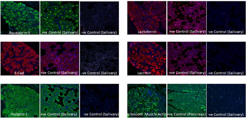
Figure 2.

Expression of lacrimal markers in ALG. Immunofluorescence imaging shows the localization of aquaporin-5, e-cad, histatin-1, lactoferrin, lacritin and α-smooth muscle actin in ALG. Sections of salivary glands were used as positive control and without primary antibodies respectively as negative control.
