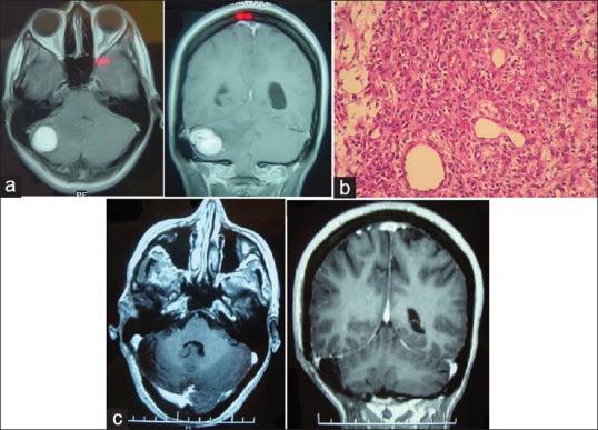
Figure 1.

(a) Preoperative brainstem MRI. T1-weighted axial and coronal images with contrast showing lesion in the right hemisphere of the cerebellum. (b) Tumor microscopy. H&E stain magnification ×40. (c) Postoperative MRI. T1-weighted axial and coronal images showing complete resection
