
Figure 1.
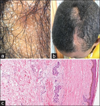
(a) Pinpoint white dots with loss of follicular openings, peripilar casts, pili torti, and areas with short regrowing hairs on trichoscopy. (b) Atrophic and depressed linear plaque of alopecia on scalp. (c) Atrophy and flattening of the rete of ridges with hyalinization and widening of dermal collagen bundles with loss of their fibrillar architecture on histology