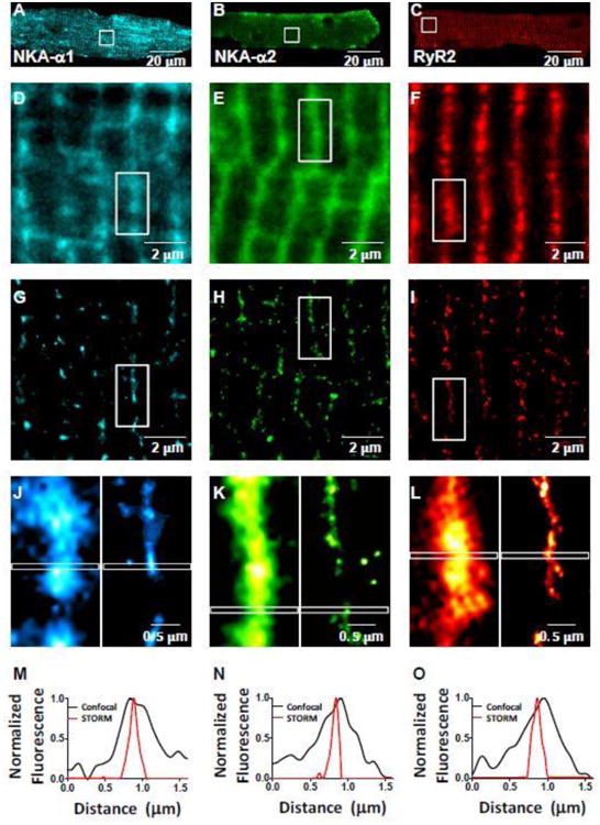
Figure 6. NKA and RyR2 STORM vs. confocal images.

A-C: Myocyte confocal images of α1 (Alexa Fluor 647) (A), α2 (Alexa Fluor 647) (B), and RyR2 (Alexa Fluor 568) (C) in mouse cardiac myocyte. D-I: Enlarged views of the box regions in A-C in confocal (D-F) and STORM (G-I) microscopy. J-L: Enlarged views of the box regions in D-I of individual z-lines (confocal left, STORM right). M-O: Plot profile of indicated regions of 1.6 μm long by 0.1 μm wide in (J-L); normalized to maximum intensity within plot profile. FWHM for STORM images were 21-26% of that in confocal images.
