
Figure 2. LEPRb-dependent STAT3 activation induces the stem cell state in LEPRb-transfected TNBC non-CSCs.
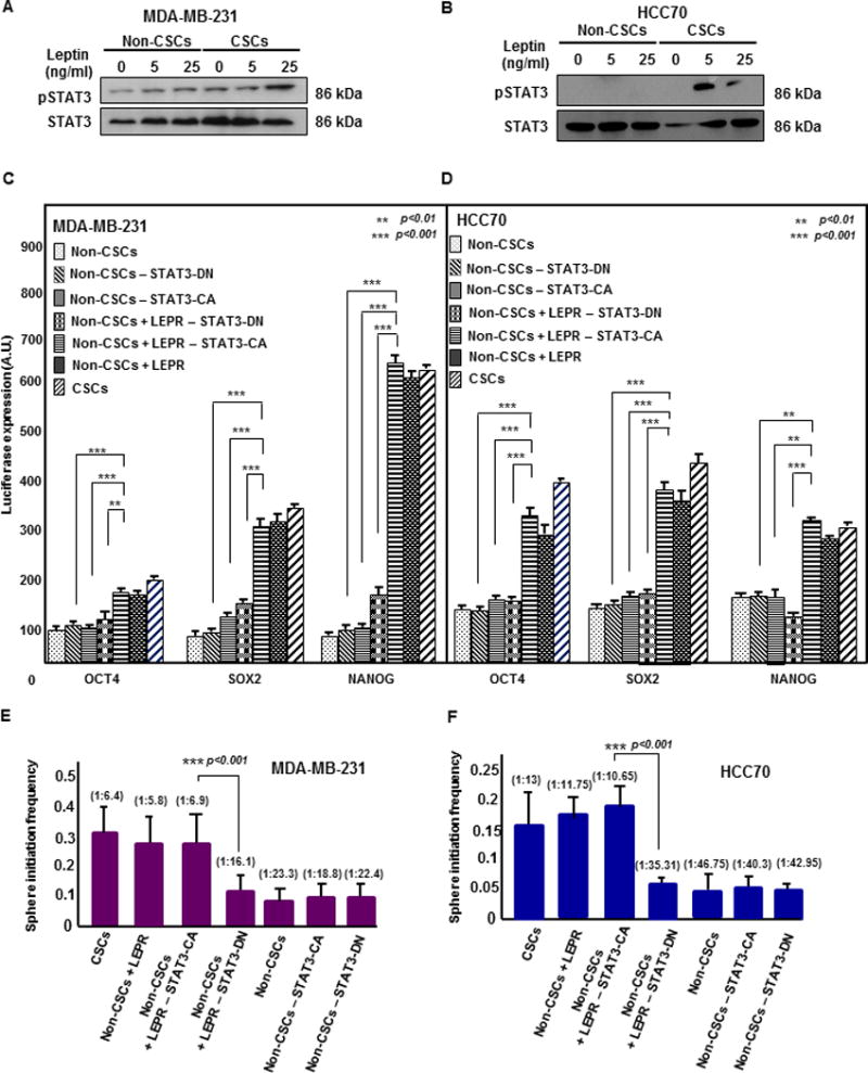
(A & B) Immunoblots of MDA-MB-231 and HCC70 CSCs (GFP+) and non-CSCs (GFP-) treated with increasing concentrations of LEP (0, 5, and 25 ng/ml) for 30 minutes were probed for pSTAT3 and total STAT3. Increased STAT3 phosphorylation was observed in LEP-treated CSCs compared with LEP-treated non-CSCs. Twenty micrograms of protein per sample was loaded into each well. (C & D) Transfection of constitutively active STAT3 (STAT3-CA) in LEPR-transfected non-CSCs (MDA-MB-231 and HCC70) increased the activity of the stem cell transcription factors NANOG, SOX2, and OCT4 as quantified by the secreted luciferase assay method. A significant increase in the transcriptional activity of all three stem cell transcription factors was observed in both LEPR-transfected non-CSC and control non-CSC groups expressing STAT3-CA compared with the corresponding dominant negative STAT3 (STAT3-DN)-expressing groups. A significant increase in transcriptional activity was also observed in STAT3-CA-expressing LEPR-transfected non-CSCs compared with STAT3-DN-expressing non-CSCs. (** p < 0.01, *** p < 0.001). (E and F) Limiting dilution analyses of STAT3-CA- and STAT3-DN-expressing LEPR-transfected non-CSCs were performed. The MDA-MB-231 and HCC70 STAT3-CA-expressing LEPR-transfected non-CSC groups showed a significant increase in stem cell frequency compared with STAT3-DN-expressing non-CSCs without LEPR overexpression (p < 0.001).