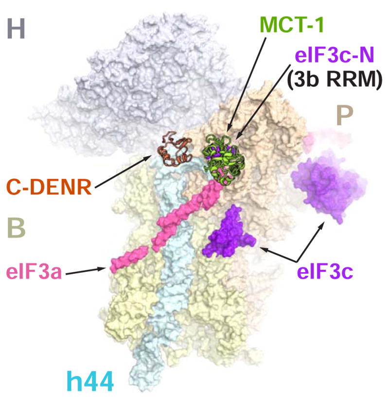
Figure 2. Binding site of the human MCT-1 on the 40S subunit overlaps with the binding site of the eIF3a and eIF3c (or eIF3b RRM) subunits of eIF3.

The crystal structure of the human 40S-DENR-MCT-1 complex was superposed with the structure of the partial yeast 48S complex in closed conformation (PDB ID 3JAP). Only eIF3a (pink) and eIF3c (purple) subunits from the yeast structure are shown. Electron density corresponding to eIF3a and eIF3c-N can also be attributed to eIF3b RRM (PDB ID 5K1H). C-DENR and MCT-1are shown in coral and green, respectively. H, head; B, body; P, platform; h44, helix 44 - domains of the 40S subunit.
