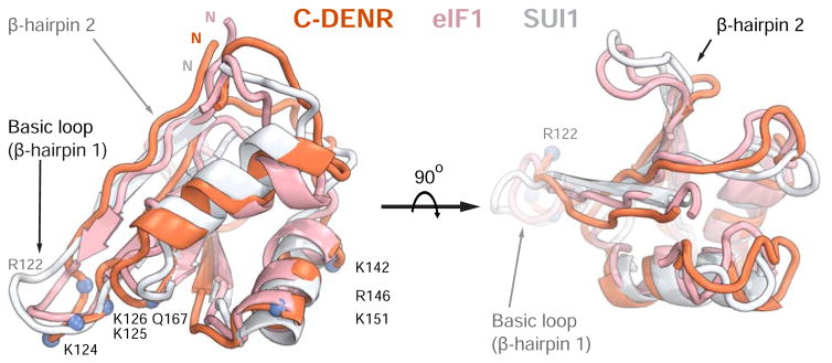
Figure 4. The structure of the C-terminal domain of human DENR.
The crystal structure of C-DENR (coral) from the 40S-DENR-MCT-1 complex was superposed with the NMR structures of the human eIF1 (pink, PDB ID 2IF1, amino acid residues 28-108) and yeast eIF1/SUI1 (white, PDB ID 2OGH, amino acid residues 24-100). The basic loop and β-hairpin 2 loop is marked by an arrow; the N-terminus of C-DENR is marked by N. Amino acid residues of C-DENR constituting the basic loop and binding site on the 18S rRNA are marked in blue.

