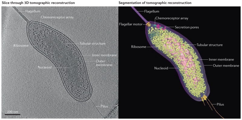
Figure 1. Example of electron cryotomography.
An intact Bdellovibrio bacteriovorus cell in standard media was plunge-frozen and imaged by electron cryotomography (ECT). The resulting tilt-series of images was reconstructed into a 3D tomogram. A slice through the reconstruction is shown (left panel), as well as a segmentation of visible cellular structures (right panel). To see the full reconstruction and segmentation, as well as fitting of crystal structures into electron microscopy densities, see Supplementary information S1 (movie).

