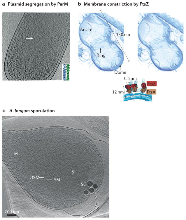
Figure 4. Division and differentiation.
Electron cryotomography (ECT) has provided mechanistic details of bacterial division, including the antiparallel doublets of ParM filaments that segregate plasmids (part a), and the copolymers of FtsZ and FtsA that can constrict membranes (part b). ECT has also revealed details of how bacterial cells differentiate into resistant spores (part c). Part a shows a slice through a tomographic reconstruction of an Escherichia coli cell expressing a high-copy-number plasmid. Inset shows a model of a ParM doublet based on a class average of 2D images of filaments formed in vitro. The top of part b is a stereoscopic image of a tomographic reconstruction of an in vitro system consisting of liposomes and FtsZ and FtsA purified from Thermotoga maritima. Below is a fit of the crystal structures of FtsZ from Staphylococcus aureus and FtsA from T. maritima into the ECT density. Part c shows a tomographic slice through an Acetonema longum cell. ISM, inner spore membrane; M, mother cell; OSM, outer spore membrane; S, spore; SG, storage granule. Part a is modified from REF. 104, Nature Publishing Group. Part b is modified from REF. 110. Part c is reproduced with permission from REF. 120, Cell Press.

