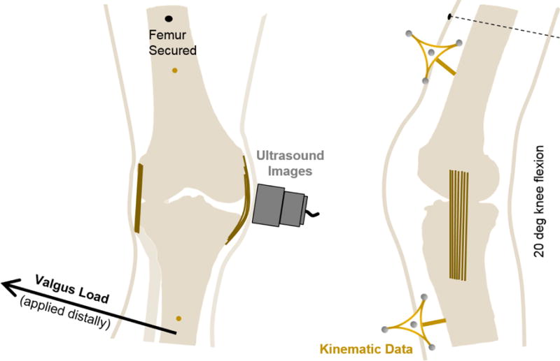
Fig. 1.

A schematic of the experimental setup. The femur was secured to a work bench at a flexion angle of approximately 20 deg. A 10 Nm valgus load was then applied to the tibia at the approximate level of the malleoli (please note that the arrow showing the application of load has been moved proximally for illustrative purposes). Simultaneously, ultrasound images were collected from the probe which was positioned over the MCL aligned along its proximal/distal axis, and motion data were collected from rigid marker frames attached to bone pins drilled through the femur and tibia.
